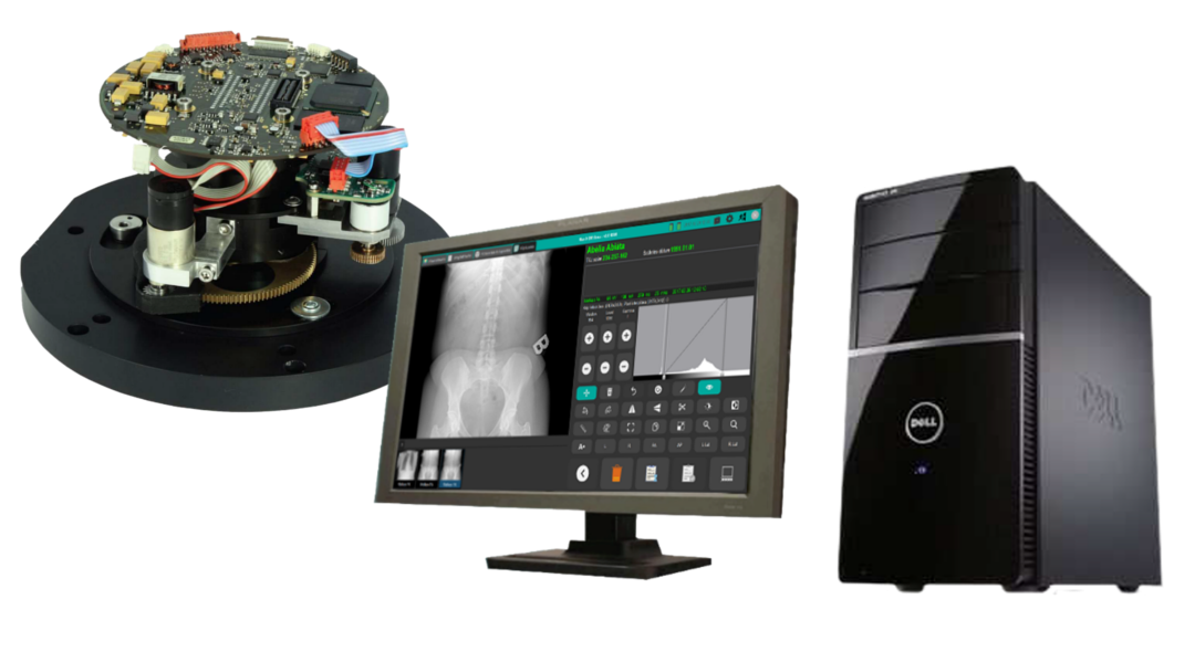
Max-X DR Spot is a high-end video spot system, composed by an image intensifier, a digital CCD camera with motorised iris, a controller PC with special imaging software, and diagnostic radiology monitors. The system is suitable for both to build new digital R/F workstations and to post-digitize of existing conventional or remote controlled R/F workstations as well.
During the R/F examination, the resulting live image on the output window of the image intensifier will be acquired and digitized by the high-resolution progressive scan CCD camera, and transferred to the workstation PC using a high-speed Ethernet cable. The life images displayed on grayscale radiology monitors can be stored on the HD of the PC in single storage, serial storage, burst storage or movie mode. The controller PC is connected to the hospital PACS as a standard DICOM modality.
In the Max-X DR Spot system, the X-ray image is created on a special low-noise and low-distortion image intensifier. This vacuum technology device specially developed for digital imaging is available in various input screen diameter and different zoom factor versions.
In case of post-digitization, the existing image intensifier having the appropriate parameters, can be kept.
The central element of the Max-X DR Spot system is the special camera-lens-iris unit, connected directly to the output window of the image intensifier. The resulting image will always be optimally projected onto the image sensor chip of the high-sensitivity and extremely low-noise CCD camera, by the specific flat lens with the built-in motorized iris control. Due to the low power consumption and excellent temperature compensation the camera works stably in the entire operating range without any additional cooling, so that high image quality is guaranteed in all modes.
Digital images will be sent to the control computer via high-speed Ethernet cable. The connection to the detector is Gigabit Ethernet, and it communicates to the PACS system through standard DICOM protocol. Perfect image quality is provided by built-in image processing services such as recursive filter with motion detection or continuous substraction of the displayed live image on a pre-stored reference image (mask). The beam parameters’ control of the related X-ray generator is provided by the configurable ABS and AEC output signals.
The operation of the Max-X DR Spot system is controlled from the integrated touch screen console located in the radiation protected control room, or from the radiology monitor cart placed in the examination room. The operator can handle patient administration, set the parameters of the X-ray generator, and control image processing from here. R/F live and stored images are also displayed here as reference.
The next patient and the associated examination can be selected at once from the worklist arrived from the PACS, but local input is also possible. If the foot switch is pressed, you can start continuous or pulse fluoro (depending on selected mode), Pressing the REC button will start single or serial storage, while pressing the EXP button will start exposure on the image receptor (conventional film cassette, CR cassette or digital flat panel detector) inserted into the spot film unit of the examination device. At the end of the examination, the images stored in the control computer can be checked and post-processed (windowing, zoom, flip, labeling) if necessary, than can be forwarded to the hospital PACS for storage and further processing (reporting, printing, archiving etc.).
The Windows based software provides wide range of image-optimization and post-processing functions. Thanks to the ergonomically designed large icons, its usage is safe and error-free in a wide range of room lightings.