Max-X DR Mammo is a full-field digital mammography system. Thanks to the outstanding image quality and high patient throughput, it is an ideal choice for both clinical diagnostical and screening applications as well. The motorized isocentric C-arm, and the motorized collimator & filter changer, set their position according to the selected examination automatically. The „smart compression” technique and ergonomically designed compressor plates ensure quick positioning and high patient comfort. Thanks to the built-in high frequency X-ray generator, high-speed X-ray tube, and high-sensitive large area flat panel detector, the system produces high-quality, high-resolution digital images with very low patient dose.
The touch screen operator console of the system functions as control surface of the HF generator as well, and is connected to the hospital PACS network as a standard DICOM modality.
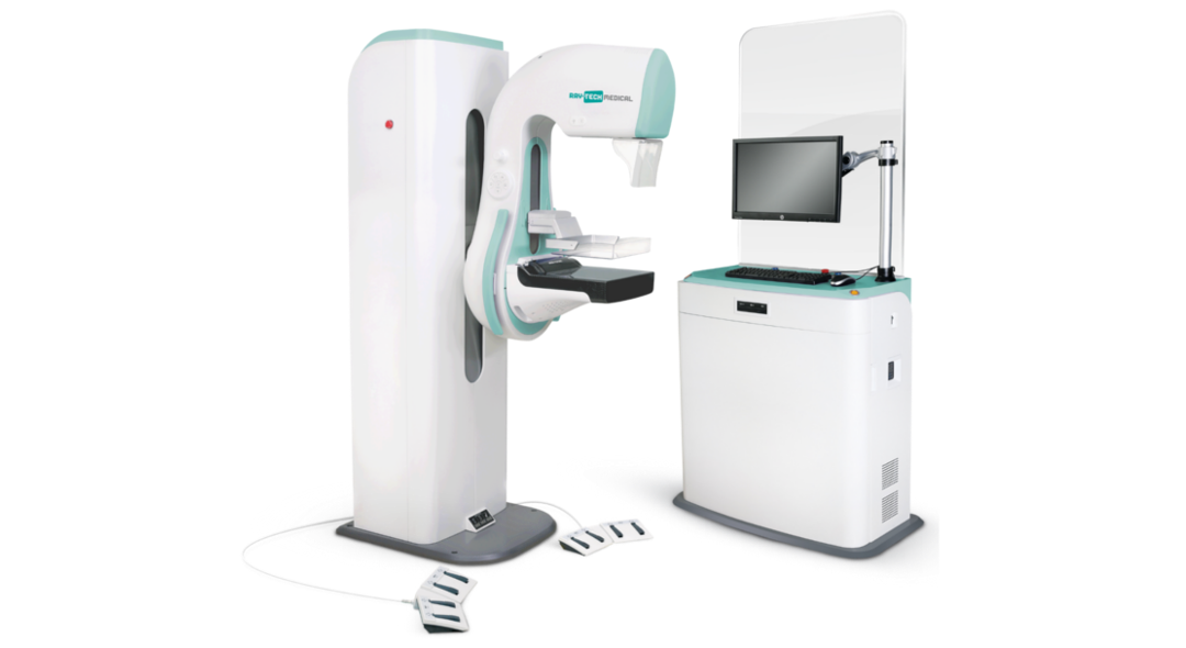
In the Max-X DR Mammo system, easy patient positioning and high-level patient comfort is provided by the design award honored, very easy-to-move, isocentric-rotated, motorized C-arm. The mechanics controlled by keys and foot pedals from both sides, is automatically set to examination position received from the DICOM worklist or set by the operator, but can also be quickly and easily changed manually. Due to the applied „smart” control, and the extremely smooth-surface, rounded compression paddles, the motorized, microprocessor controlled compression unit with automatic compression force allows gentle positioning in all radiographic modes. The precise and optimal patient positioning is supported by the wide range of compressor paddles with lateral shift. C-arm angle, compression force and thickness can be seen on large-size digital displays, so these parameters can be quickly checked and adjusted both from the device and the control console.
For spot X-rays, the system includes a two-stage, geometric magnifying attachment as well. Its connection is immediately detected by the detector that adjusts system settings (focus, collimator, tube current) accordingly.
In the Max-X DR Mammo system, X-ray is provided by a built-in, microprocessor controlled, high frequency X-ray generator and its associated rotating anode fine focus X-ray tube. The motorized collimator and filter changer will automatically adjust to the selected exam, without intervention by the operator.
The generator control is integrated to the common touch-screen control panel of the imaging system. Optimal image quality is guaranteed by the built-in AEC dose control and the removable fine grid.
The heart of the Max-X DR Mammo system is the 24x30 cm high resolution direct digital flat panel detector. This semiconductor image receptor, specially designed for mammography purposes is capable of creating high quality fine X-ray images with very low patient dose. Within 10 seconds after exposure, the image appears on the touch screen monitor, and on the interpretation monitor, as an option.
The Max-X DR Mammo system is controlled from a large, integrated touch screen console completed with a radiation shielding lead glass. All functions are accessible here: waiting list handling, patient administration, study selection, generator control, and remote control of mechanical movements. It displays the preview image after exposure, and when the operator checked, it can be sent to the connected PACS for storage and processing (interpretation, printing, archiving etc). In automatic mode the software sets the optimal generator parameters according to the selected examination protocol and measured data. The built-in software supports a wide range of image post-processing functions. The ergonomic design and large icons ensure reliable control in any environment.
In case of stand-alone installation or temporary connection error to the HIS/PACS system, local patient administration, study selection, reporting and printing are available as well.