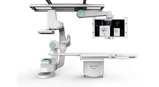
Max-X DR Angio W is a single-plane image guided DR system for cardiovascular and interventional imaging. Ideally suitable for cardiac, peripheral, abdominal and neural interventional examination and treatment. The floor-mounted fully-motorized C-arm can be set to any preprogrammed or freely selected imaging position quick and safe. The motorized 6-way cardiovascular patient table with extra large movement range allows easy access to patient in any examination procedure. The high resolution large image area dynamic flat panel detector provides cristal clear images with wery low patient dosage. The large all-in-one monitor mounted on ceiling rails can display all relevant information along with the actual X-ray images (like ECG and breathing curves, blood pressure and oxigen saturation data, images from CT/US and other modalities etc.). Wide range of selectable image processing software tools help the visualization and right diagnose. The system is connected to the hospital PACS as a standard DICOM modality.
The imaging components of the Max-X DR Angio W system are assembled on a floor-mounted fully-motorized C-arm, can be set to any preprogrammed or freely selected imaging position quick and safe. The X-ray is generated by the 0.3/1.0mm fine focal spot dual cooling X-ray tube located on one end of the C-arm, while the digital image is acquired by the dynamic flat panel detector located on the other end of the C-arm. The high resolution large image area detector provides cristal clear radiography and fluoroscopy images with wery low patient dosage. The automatic patient conturing provided by the real-time motorized detector position-correction system assures optimal image quality with maximal patient safety in any examination procedure.
The easy positioning of the patient in the Max-X DR Angio W system is assured by an integrated motorized cardiovascular patient table. The 6 way movement table has carbonfiber tabletop and flat surface with low X-ray attenuation. The 2m longitudinal travel range ensures head to toe eamination without patient moving, and no C-arm moving needed when switching examination from cardiology to neurology. The attached table-side control panel provides quick and easy control for all major components: table movement, C-arm movement, collimator control, image play-back control, SID control, etc.
All major information in the Max-X DR Angio W system is displayed on a ceiling suspended full-color large size medical grade monitor. It can display all relevant information along with the actual X-ray images (like ECG and breathing curves, blood pressure and oxigen saturation data, images from CT/US and other modalities etc.). The screen layout and the related content can be set according to the performed procedures, and can be controllled real-time directly at table side.
In the Max-X DR Angio W system X-ray tube is powered by a modern, microprocessor controlled, 100kW high frequency X-ray generator. The generator control is integrated to the common touch-screen control panel of the imaging system. Optimal image quality is guaranteed by the built-in ABS and AEC dose control. The supervisor program monitors the system status and any operator intervention all the time - thus effectively preventing any setting or events that could cause damage or overload to any component of the system.
The Max-X DR Angio W system is controlled from an integrated multi-monitor central operator station. All functions and information are accessible here: worklist handling, patient administration, study selection, generator control, mechanical positioning. It displays the actual live and recorded X-ray images along with all relevant other information (like ECG and breathing curves, blood pressure and oxigen saturation data, images from CT/US and other modalities etc.). Wide range of selectable image processing software tools help the visualization and right diagnose (angiography, DSA, RDSA, 3D-CT, 3D-DSA tools). The system is connected to the hospital PACS as a standard DICOM modality.
Wide range of connectable external devices